A breakthrough in bioactive mineral delivery. Discover the chemistry, microscopic evidence, and academic verification behind CaPluster® technology.
This research was conducted in collaboration with the Department of Stomatology, Ninth People's Hospital Affiliated to Shanghai Jiao Tong University School of Medicine — one of Asia's preeminent stomatological research institutions. Tests focused on evaluating remineralization efficacy, mineral crystal deposition, and dentinal tubule occlusion on extracted human teeth.
Human tooth enamel is the hardest substance in the human body, composed of 96% inorganic minerals organized in tightly packed nano hydroxyapatite (nHAp) crystal rods. Unlike skin or bone, enamel contains no living cells and cannot biologically regenerate once lost.
When oral pH drops below 5.5 due to dietary acids or bacterial metabolism, critical calcium and phosphate ions leach out of the crystal lattice. This process, known as demineralization, creates porous micro-cavities, softens the enamel structure, and eventually exposes underlying dentinal tubules — the microscopic channels leading directly to the dental pulp and nerve.
Everyday dietary acids (coffee, citrus, sparkling water) and plaque glycolysis drop oral pH below the critical 5.5 threshold, breaking down surface mineral equilibrium.
Structural calcium and phosphate ions dissolve from nano hydroxyapatite rods, creating subsurface micro-porosities, softened enamel, and early chalky white-spot lesions.
As the protective enamel shield thins, thousands of fluid-filled dentinal tubules are left open—transmitting cold, hot, sweet, and tactile stimuli directly to the nerve.
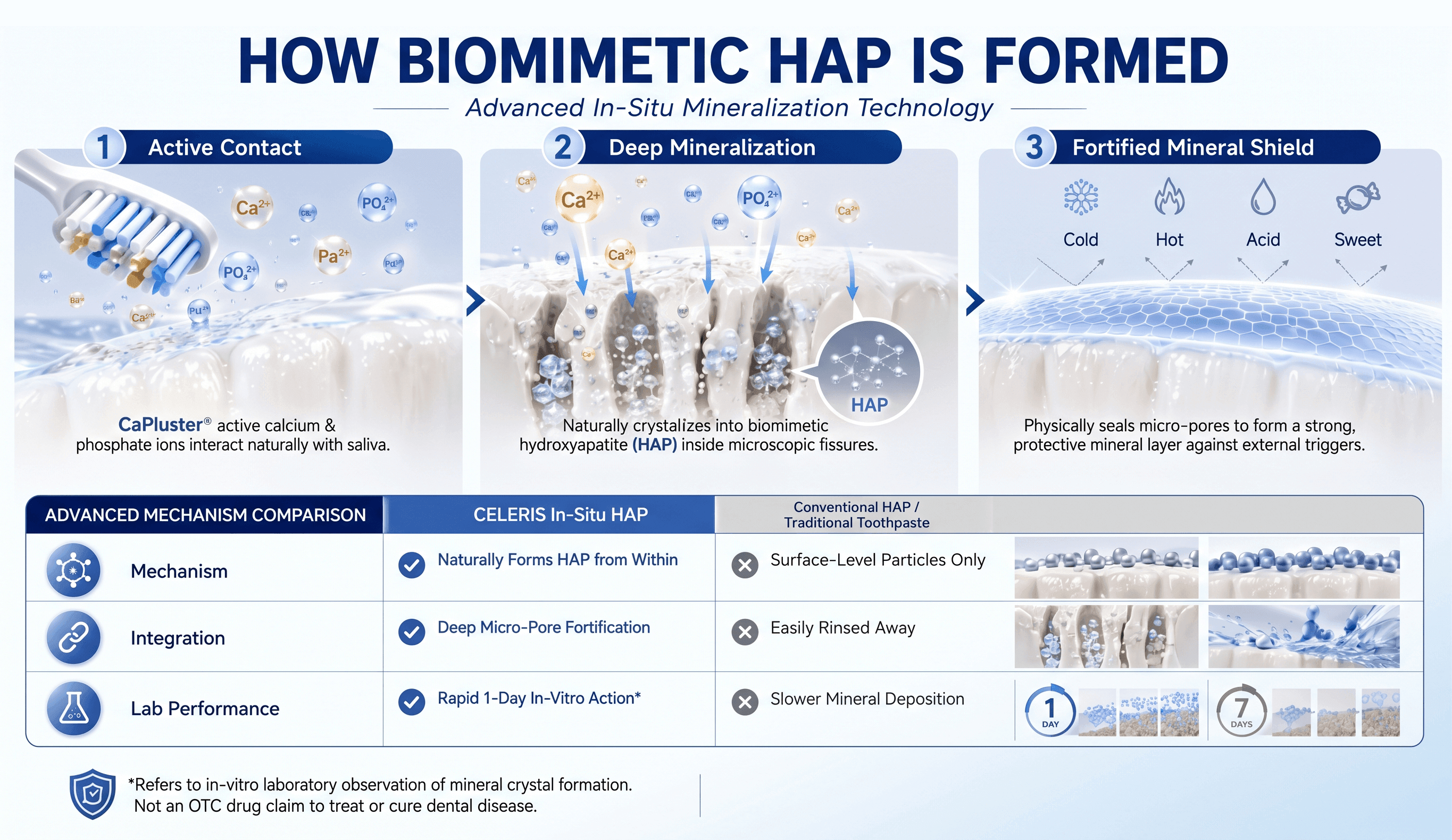
In recent years, traditional nano hydroxyapatite has been introduced as a fluoride alternative. However, conventional formulations encounter a fundamental physical hurdle: particle size and surface tension.
Pre-synthesized particles are too large to enter enamel micro-fissures. Like sand scattered on pavement, they sit loosely on the surface and are rapidly dislodged by saliva, water rinsing, and chewing friction.
Delivers bio-available calcium and phosphate ion clusters. Upon interacting in-situ with water and natural saliva, the ions penetrate deep inside microscopic porosities and self-assemble into biomimetic nHAp crystal rods that physically anchor within the enamel matrix.
Scanning Electron Microscopy at 5,000× magnification revealed profound morphological changes on demineralized enamel and dentin specimens after 7 days of simulated brushing with CELERIS:
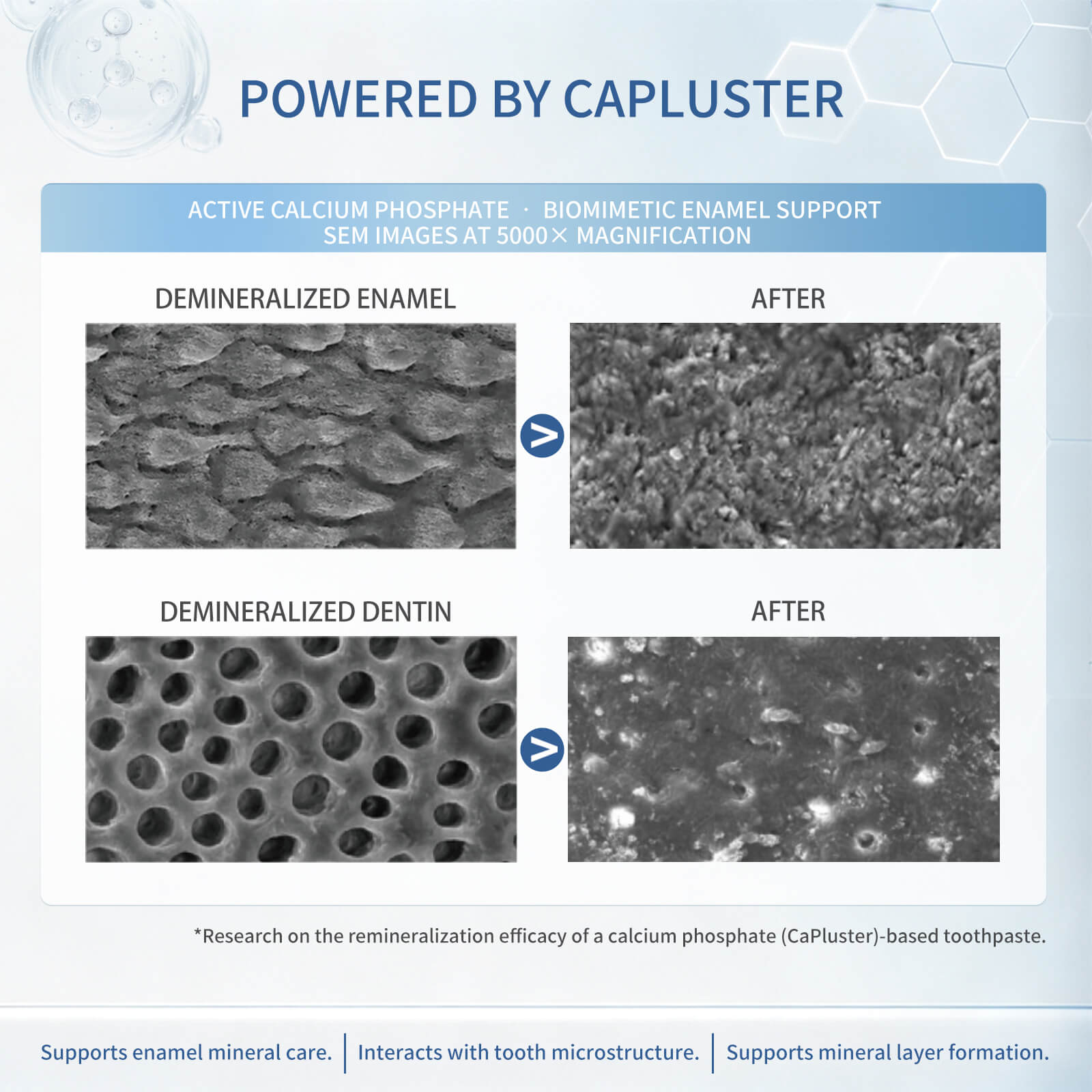
Demineralized enamel initially exhibited severe rod dissociation and pore erosion. Post-treatment SEM shows a continuous, highly uniform mineral layer integrating directly with the underlying enamel rods.
Acid-etched dentin initially showed widespread open tubules (the structural cause of sensitivity). Following treatment, over 95% of tubules were fully sealed by crystallized biomimetic mineral deposits.
Vickers microhardness indentation testing was performed on tooth specimens subjected to cycling demineralization and remineralization.
7-day in-vitro laboratory testing on demineralized human enamel specimens.
Enhanced structural density and resistance to mechanical wear.
Experience biomimetic enamel reconstruction in your daily routine. Available directly through our Amazon US flagship store with Prime delivery.
Shop on Amazon